Wednesday, 10 August 2022

Not so FAST? How a study on lung ultrasound got it wrong…


This study out of UC San Diego set out to determine the accuracy of lung ultrasound vs. supine portable chest x-ray for the detection of traumatic pneumothorax.

Yes, I also thought this research questions had been suitably answered.

In 2020, a CochraneReview included 13 prospective paired comparative accuracy studies and concluded that ultrasound was about 90% sensitive vs. 50% for a chest x-ray. Both are highly specific.

Regardless of the consistent evidence, let’s consider face validity. Anyone who is competent in lung ultrasound knows that ultrasound is much better at finding pneumothorax. There is no planet in the solar system where a supine portable chest x-ray is going to be more sensitive than ultrasound.

But this study concludes the opposite… chest x-ray was better!

Huh? How could this be?

This was a retrospective registry review. Retrospective data is often poor quality. Registry data is even worse. Garbage in... garbage out. Ironically, this study would never have been included in the Cochrane review.

What is the probability that this single study with worse quality evidence is correct and all the consistent higher quality studies are wrong?

Yes, the probability is close to zero.

But where did this study really go wrong?

I think the authors answered the question themselves, “… in a non-study environment, such as ours, the pleural views were not universally performed with the same rigor as in a prospective study designed specifically to detect pneumothorax.”

I think this is probably the answer.

There are many other problems with this study, but I’m limited by word count. So briefly, the  methods for assessment of diagnostic accuracy of a test were performed backwards. They used the wrong probes for lung ultrasound. They used the wrong clinicians to perform and interpret the ultrasound. That was only to name a few...

As expected, there were many concerned letters to the editor. Even one from Dr Giovanni Volpicelli who is the world leader and pioneer of lung ultrasound. But the authors stuck with the strategy of “never admit you are wrong” and doubled-down in their conclusions.

A bit sad really...


Covering:

Santorelli JE, Chau H, Godat L, et al. Not so FAST- Chest ultrasound underdiagnoses traumatic pneumothorax. J Trauma Acute Care Surg.2022;92:44-8.[link to article]


Friday, 3 June 2022

Nonoperative or Surgical Treatment of Acute Achilles’ Tendon Rupture


Why
do emergency clinicians need to know about this paper? Afterall, we will not be making treatment decisions about to operate or not…

Two reasons.

First, we need to know the “next step” and be able to inform our patients of what the treatment options are likely to be.

Second, this was an RCT published in the New England Journal of Medicine. This is the highest impact medical journal on the planet. Papers published here set the guidelines about how we practice medicine.

These authors from Norway randomized 554 patients with acute Achilles tendon rupture to one of three treatment arms: conservative, open repair, or minimally invasive surgery.

The primary outcome was the change in the Achilles tendon Total Rupture Score at 12 months. They also looked at incidence of tendon re-rupture.

They standardized the aftercare & rehabilitation in all groups. They required casts and weight bearing as tolerated using an ankle-food orthosis with heel wedges for 6-8 weeks. They did not report on earlier return to functional status between groups.

Results?

At 12 months, there was no difference in the rupture score. As expected, there were more re-ruptures in the conservative vs. surgical arms (6.2% vs. 0.6%). And there were more nerve injuries in the surgical groups.

All studies have limitations and this one is no exception. An RCT is only as good as the measuring tool of its primary outcome. I’m not sure what to make of the rupture score and self-reporting may have led to bias.

What are we to make of this?

In the long term, there probably is no big difference between conservative vs. surgical management.  

Nevertheless, there is probably enough wiggle room with the interpretation of this study for orthopedists to continue doing whatever they like. Conservative seems fine in the long run, but there are higher rates of re-rupture. Surgical treatment is about the same but comes with the potential for nerve injury and infection.

Seems like we are right back where we started!


Covering:

Myhrvold S, Brouwer E, Andresen T, et al. Nonoperative or Surgical Treatment of Acute Achilles’ Tendon Rupture. N Engl J Med 2022;386:1409-20. [link to article]

Risk of delayed intracranial hemorrhage after an initial negative CT in patients on DOACs with mild traumatic brain injury


To identify delayed hemorrhage, some clinicians will repeat a head CT in patients on NOAC’s with mild traumatic brian injury (TBI) after the initial head CT is negative.

How common is delayed hemorrhage with DOAC’s?

Hmmm….

These authors from Italy performed a retrospective multicentre observational study. They found 1426 patients taking NOAC’s who were evaluated in the ED for mild TBI (GCS 14-15). Of these 916 (68%) underwent 24 hours of observation and a repeat CT after the initial one was negative.

In the end, only 14 (1.5%) patients had delayed hemorrhage found on repeat CT. None of these patients had neurosurgery or died (but I presume they had their DOAC’s withheld).

Another interesting aspect… no delayed hemorrhages were found in patients who had the initial head CT greater than 8 hours after injury.

The findings of this study are similar with others looking at warfarin. Consistency in the literature is comforting.

So, we have a pretty simple message. Routine delayed head CT is not necessary. But 1.5% is not zero… so we still need to be a little careful.

And another question… is it DOAC’s or NOAC’s?


Covering

Turcato G, Cipriano A, Zaboli A, et al. Risk of delayed intracranial haemorrhage after an initial negative CT in patients on DOACs with mild traumatic brain injury. Am J Emerg Med. 2022;53:185-189. [link to article]

Single dose phenobarbital for the outpatient treatment of alcohol withdrawal... interesting


Phenobarbital has some ideal properties for the treatment of alcohol withdrawal. In particular, it has a long half life (about 100 hours) allowing for a single dose without the requirement for prescriptions.

However, there are some genuine safety concerns especially if they are mixed with other drugs or alcohol. And there is little high-quality evidence to support it’s use in patients being sent home from the ED.

This is a retrospective chart review of patients discharged from a single ED in California over a three-year period with a discharge diagnosis of alcohol withdrawal. Patients were stratified into three categories; those who got benzo’s, benzo’s plus phenobarbital or just phenobarbital alone.

The primary outcome was return ED encounter within 3 days of the index visit. (Yes, this is an odd primary outcome, but it was chosen for pragmatic purposes as below...)

470 patients were included. 235 got benzo’s, 133 got phenobarbital, and 102 got the combination special.

Results?

Treatment with phenobarbital (alone or in combination) was associated with a decreased odds ratio of a return ED visit within 3 days. (AOR 0.45 95% CI 0.23, 0.88  and AOR 0.33 95% CI 0.15,0.74 respectively).

The cynic in me wonders if this is because phenobarb patients died… (But this was probably not the case as the author were eventually able to account for most patients)

It is interesting to note that patients who received phenobarbital got much more GABA agonist medication than those that simply got benzodiazepines. Four times as much was given if you adjust for drug equivalence. Perhaps this is why they were less likely to return?

Unfortunately, this paper does not address the big question of safety. It would take a much larger study with more robust methodology (that avoids loss to follow-up) to answer this. And such a study would be very difficult to undertake as this cohort of patients are unreliable and not likely to be compliant with study protocols etc.

What’s the take home message?

Phenobarbital for the outpatient treatment of alcohol withdrawal is tempting, but there is arguably not enough evidence to support its routine use.


Covering:

Lebin A, Mudan A, Murphy CE, et al. Return Encounters in Emergency Department Patients Treated with Phenobarbital Versus Benzodiazepines for Alcohol Withdrawal. J Med Tox. 2022;18:4-10.  [link to full text of article]


Thursday, 2 June 2022

Risk for recurrent VTE in patients with subsegmental PE managed without anticoagulation- A prospective study


Modern imaging technologies have put a virtual microscope into the human body and can find smaller and less significant disease. Our treatment of these conditions often falls under old recommendations meant for more substantial illness. Can treatment of mild disease result in more harm than benefit?

This question is particularly true when it comes to subsegmental PE. Does it require anticoagulation?

These researchers found 266 patients with low-risk subsegmental PE and referred them to a thrombosis clinic for management. The patients underwent lower extremity ultrasound to exclude DVT at the time of diagnosis and one week later. Patients without DVT had anticoagulation withheld and they were followed up for 90 days looking for evidence of recurrent VTE.

In the end, 8  (3% 95% CI, 1.6%-6.1%) out of the 266 patients developed VTE. The authors report higher rates in some subgroups but this analysis is not really justified given the low number of recurrences. 

There are some important limitations to remember. These patients were low risk. They did not have cancer nor prior VTE. They all had repeated leg ultrasound to exclude DVT. Long term rates beyond 90 days were not assessed. In addition, false positive CTPA's may have skewed the results towards lower rates of recurrent disease. 

What are we to conclude?

Recurrent VTE in low-risk patients with subsegmental PE is pretty low. This may be a good time for shared decision making.


Covering:

Le Gal G, Kovacs M, Bertoletti L, et al. Risk for Recurrent Venous Thromboembolism in Patients With Subsegmental Pulmonary Embolism Managed Without Anticoagulation. Ann Intern Med. 2022;175:29-35 [link to article]

Thursday, 14 April 2022

Cannabinoid hyperemesis syndrome: A 6-year audit of adult presentations to a single ED in Melbourne


I'll preface everything by stating this paper is not high science.

Sorry…

These authors performed a retrospective chart review on adult patients who they thought had cannabinoid hyperemesis syndrome (CHS) in their single ED in northern Melbourne.

Obviously, the diagnosis of CHS is based on clinical findings. There is no blood test for CHS and no agreed formal diagnostic criteria. So, who knows if they identified all patients with CHS or if the ones they diagnosed truly had it?

Nevertheless, there might be a few useful pieces of information… so let’s continue on our magic carpet ride.

142 patients were included. 77 were unique presentations and the rest were frequent visitors.

I’m not going to report their clinical features as this formed part of how they identified patients in the first place… kind of an incorporation bias. (Suffice to say, they were vomiting a lot.) Plus, the retrospective nature of the data means a lot of things were probably not recorded (i.e. relief from hot shower was only written down 11% of the time).

More objectively, I was interested to see that most had elevated white blood cell counts and lactates. Median WBC was 14 (IQR 11.2-16.8) and lactate was 1.95 (IQR 1.4-2.9)

These doctors also liked droperidol and the authors go on to state, “Response to droperidol (or haloperidol) as previously reported may also guide clinicians in their determination as to whether CHS should be considered.”

Arguably the best part of the paper is their proposed CHUNDER score to aid in the diagnosis of CHS. This mnemonic stands for, “Cyclical vomiting, History of regular cannabis, Under fifty years old, Normal lipase, Diagnosis of exclusion, Elevation of CRP<50, and Reduction in symptoms after droperidol.

I’m chunder struck…


Covering:

Rotella J, Ferretti O, Raisi E, et al. Cannabinoid hyperemesis syndrome: A-year audit of adult presentations to an urban district hospital. 2022 Feb 23. doi: 10.1111/1742-6723.13944 [link to article]


Association of IV Contrast with Renal Function- A Regression Discontinuity Analysis


IV Contrast has been around for decades. How come we still do not know whether its causes renal damage!

Well, today is your lucky day…

These researchers conducted a randomized double-blind placebo-controlled trial of IV contrast in 2000 patients with borderline renal function to determine if there was any meaningful worsening in patient-oriented outcomes.

Just kidding!

There are limits in conducting clinical trials… the biggest of which boils down to ethics and feasibility. Sometimes, an RCT is not going to happen. So, we are stuck with observational studies which have the inherent trouble in dealing with confounding and attributing causality.

In an attempt to overcome these limitations, these researchers performed a regression discontinuity design (RDD).

Huh?!?

The RDD relies upon the existence of a continuous variable (i.e. D-dimer) for which there is a cut-off that determines treatment or investigation. Patients on either side of the cut off have much different probabilities of investigation but probably are reasonably matched for confounding factors.

Pretty clever…

These researchers included adult patients who had a D-dimer test during an ED visit over a 5-year period in the province of Alberta, Canada. They compared the group that had elevated D-dimers vs. those that were normal. The primary outcome was long term renal function (up to six months from ED visit).

Results?

150,028 patients met inclusion criteria. Mean baseline eGFR was 86.

There was no evidence of renal damage with a change of eGFR of only 0.4 (95%CI -4.9 to 4.0) up to 6 six months later.

The authors conclude; 

“To our knowledge, this study provides the strongest evidence to date that intravenous contrast is not associated with significant kidney injury…”

This overall conclusion is correct. But... we are not worried about patients with near normal renal function to start with. We are much more interested in the cohort that has borderline function.

They tried a subgroup analysis by looking at patients with eGFR <45 but state that the analysis was hindered by missing data and small numbers. This is unfortunate.

What should we conclude?

Overall, the modern IV contrast agents probably don’t do much to kidneys. But we are not entirely sure in the cohort with more severe baseline renal impairment.


Covering:

Goulden R, Rowe B, Abrahamowicz M, et al. Association of Intravenous Radiocontrast with Kidney Function. A Regression Discontinuity Analysis. JAMA Intern Med. 2021;181:767-774. [Link to article]


Saturday, 9 April 2022

Rapid agitation control with ketamine in the ED: A Randomized Controlled Trial


Agitated patient tearing apart the ED...? Which IM medication do you give?

These authors from a single ED in Vancouver randomized patients to either ketamine 5mg/kg IM vs. midazolam 5mg plus haloperidol 5mg IM.

Their primary outcome was time to adequate sedation as measured on a validated agitation score.  

(They tried to blind the dose but it’s hard to blind ketamine when we all know what it looks like. Either way, this was probably only a minor source of potential bias.)

Results?

Enrolment was cut a bit short at 80 patients due to the start a supposed global pandemic.

Median time to adequate sedation was 6 minutes in the ketamine group vs. 15 minutes in those randomized to midazolam/haloperidol. 

So, ketamine is the clear winner?!?!

There are two major reasons why this conclusion is wrong.

Firstly, they compared a grizzly bear dose of ketamine to a low dose of midazolam & haloperidol. For what it is worth, I usually start with midazolam 10mg and droperidol 10mg (unless there is reason to start lower… elderly, co-morbid, etc.) Droperidol works faster than haloperidol.

Secondly, they asked the wrong research question. I care less about how quickly someone is sedated and care much more about how well it works overall.  I’m sure a dose of IM sux would work pretty quick too. Or even a bullet to the head would have an immediate calming effect (but would be associated with excess mortality.)

How often did the ketamine group require re-sedation? How well did the patients wake up? Lots of adults complain about side effects of ketamine including dysphoria to emergence phenomenon. But what could go wrong with giving an agitated patient a medication similar to PCP?

To be fair to the researchers, it is difficult to objectively measure, “how well did it work?” It's complicated and there is some subjectivity in this question. Time to sedation is much easier to measure… but just because we can measure something accurately does not necessarily mean it is important. (This kind of reminds me of the streetlight effect).

In short summary, I’m willing to believe a big slug of ketamine will cause someone to drop quickly. But is this the best thing for the patient? I doubt it.


Covering:

Barbic D, Andolfatto G, Grunau, et al. Rapid Agitation Control with Ketamine in the ED- A Blinded, Randomized Controlled Trial. Ann Emerg Med. 2021;78:788-795 [link to full text article]


The erector spinae plane block for acute pain management in ED patients with rib fractures


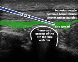
The erector spinae plane block provides analgesia for rib fractures. It is similar to 3 in 1 blocks that we are comfortable performing for femoral neck fractures. It requires ultrasound guidance and high volume (30-40ml) of dilute anesthetic to allow the medication to spread below a fascial plane. 

For a decent video demonstrating the block, check out: https://www.youtube.com/watch?v=O9RB0K7f8pM

Interestingly, the block was only just described in the anesthesia literature in 2016 but it has already become quite the rage.

Why? It is simple, effective and the incidence of complications is felt to be low.

However, it looks like it hasn’t really made its way into the Emergency Department.

Enter two Canadian emergency doctors who published a case series of 9 patients. Ok… a pilot study and not great science.

The mean reduction in NRS pain scores went from about 10 to 3.5. So, it seemed to work pretty darn well. Of course, they cannot make any claims about safety, but other literature has somewhat addressed this.

The authors appropriate state that more research is needed but they “feel that the erector spinae plane block should be considered in patients with acute rib fracture pain as a method of pain control in multi-modal analgesia.”

I think this is probably a fair conclusion. We perform a multitude of blocks in the ED. With appropriate training, this can probably be added to our armamentarium.


Covering:

Surdhar I, Jelic T. The erector spinae plane block for acute pain management in emergency department patients with rib fractures. Can J Emerg Med. 2022;24:50-54. [link to article]